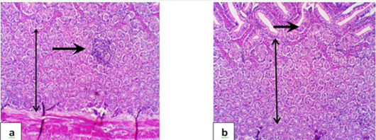
Figure 5:
Representative photomicrograph of gander intestine in G2 showing focal of mild cellular infiltrates (a), villus fusion with thickness of villous lamina epithelialis mucosa, extensive numerous intestinal crypt proliferation, and few cellular infiltrates. (H & E, x100).