
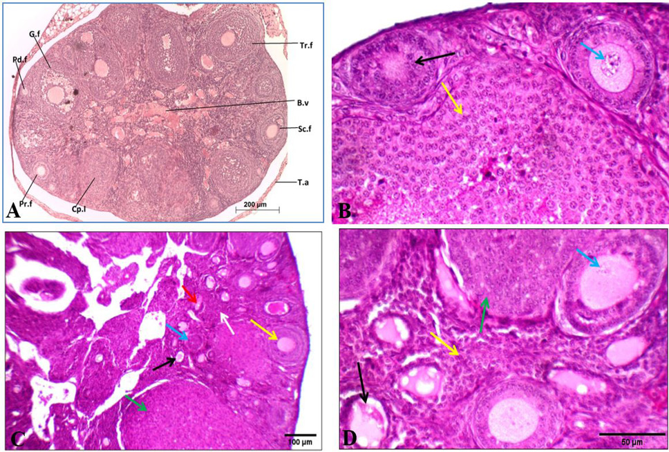
Effect of oral administration of exogenous testosterone on ovary of albino mouse. A shows histological structure of transverse section of the ovary of control female albino mice showing different stages of follicles. B.v: blood vessel, Cp.l: corpus luteum Pd. f: Primordial follicle, Pr. f: primary follicle, Sc. f: secondary follicle, G.f: Graffian follicle, T.a: tunica albuginea, Tr.f: tertiary follicle. B shows control ovary showing primary (black arrow) and secondary follicles with average oocyte (blue arrow), and corpus luteum (yellow arrow). C shows primordial (black arrow), primary (blue arrow), secondary follicles (yellow arrow), and multiple corpora lutea (green arrow) in cellular stroma (white arrow). D shows ovary from treated mouse showing primordial (black arrow) and secondary follicles with degenerated oocyte (blue arrow), and multiple corpora lutea (green arrows) in cellular stroma (yellow arrow). Stain: H and E. Magnification: A: 100X; B: 400X; C: 200X; D: 400X