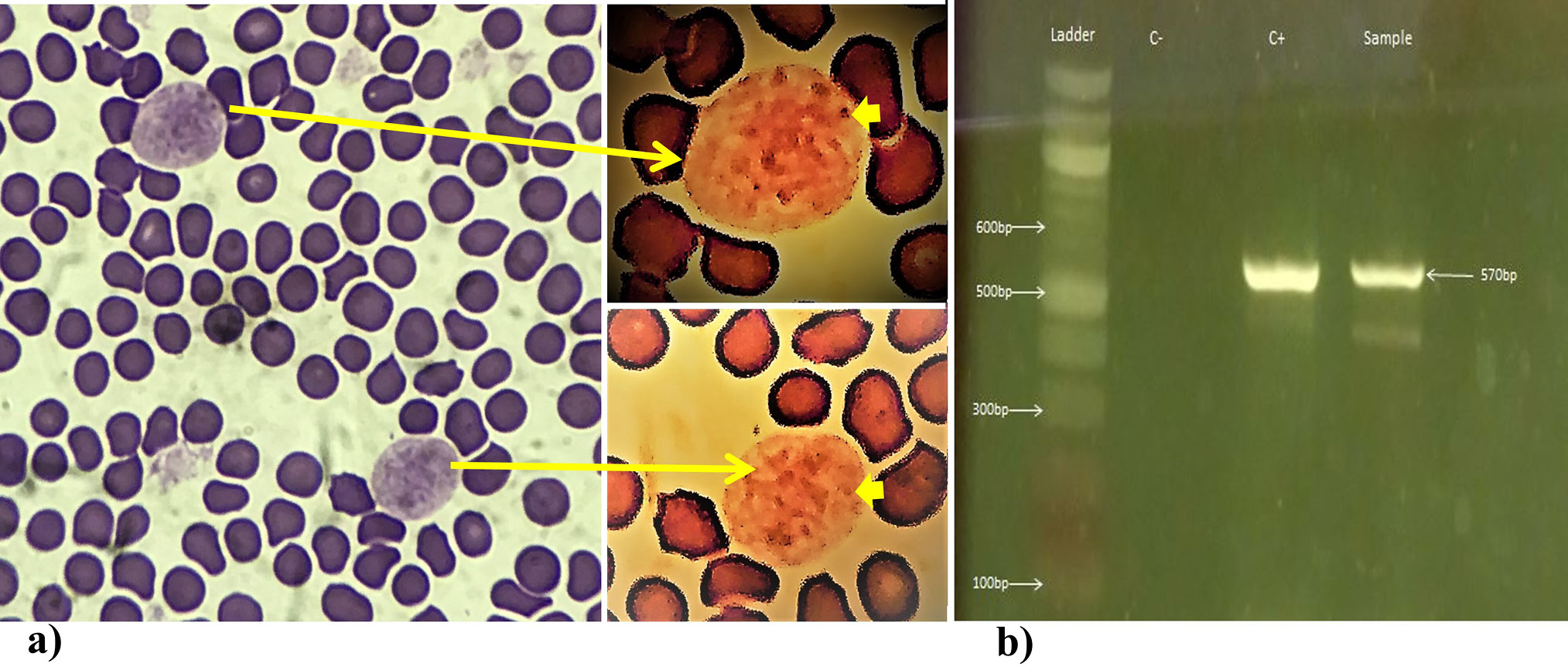
Fig. 1.
(a) Microscopic image captured with an i-Phone-7 from stained blood smear of tiger blood sample examined at 1000x magnification in an Olympus CX-21 microscope. The inset contains images of two monocytes that were edited with photos editor of windows, with a Zeke Filter on it, for a clearer view of the multiple intracellular Leishmanial amastigotes (Thick yellow arrow pointed to one of those) that actually helped in diagnosis. (b) An image of agarose gel indicating a band at the size expected for L. infantum detection in a tiger blood. C+ and C- represent positive and negative controls, respectively.