
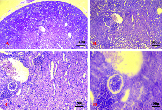
Figure 11:
A, B, C & D: Shows the kidney section in group 5 at 5th week revealing severe degeneration and necrosis of glomeruli accompanied by widening of the glomerular wall and damaged renal tubules. Aggregations of immature cancer cell and heavily invaded the renal parenchyma lead to loss of normal histological features. (A. X 4, B. X10, C.X20, D. X40). (H&E).