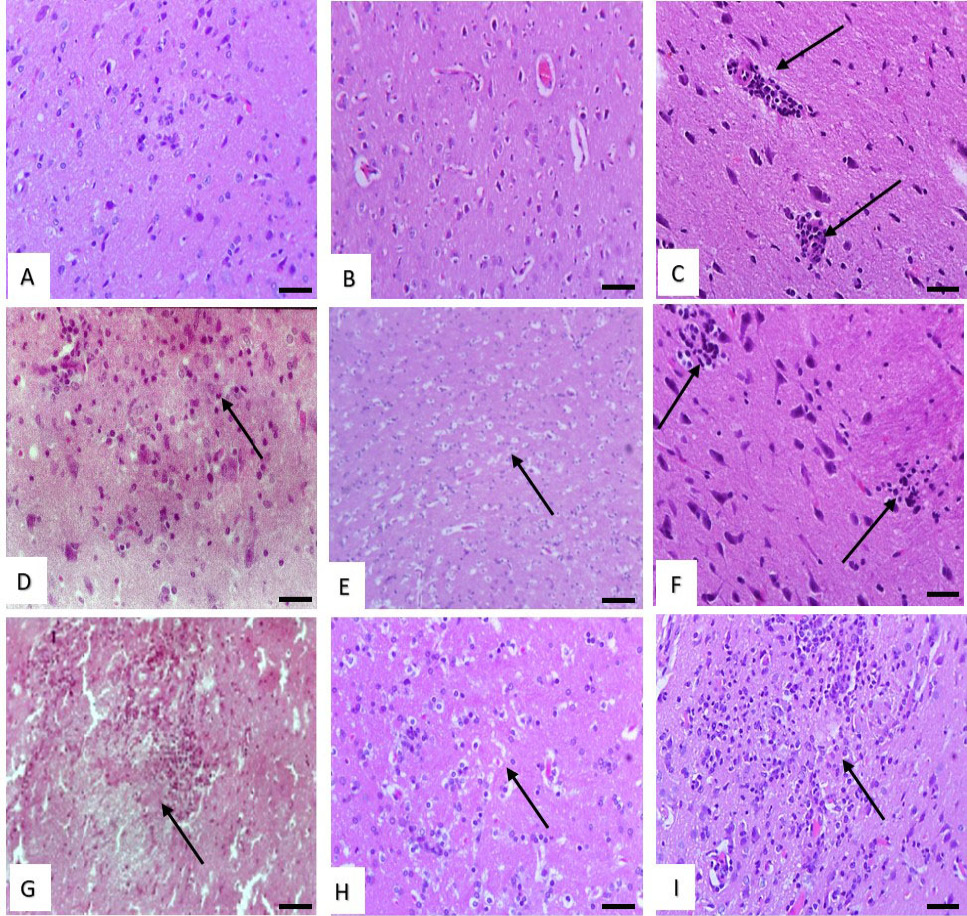
Histological structure of rat brain. (A and B) Effect of low dose of malathion and metalaxyl showing normal structure. (C) Effect of low dose of cymoxanil showing slight perivascular lymphocytes infiltration (arrows). (D) Effect of with medium dose of malathion showing neuronal cell degeneration (arrow). (E) Effect of medium dose of metalaxyl showing slight spongiosis (arrow). (F) Effect of medium dose of cymoxanil showing focal collections of glial cells forming Babe’s bodies (arrows). (G) Effect of high dose of malathion showing focal cerebral malacia (arrow). (H) Effect of high dose of metalaxyl showing cerebral spongiosis and gliosis (arrow). (I) Effect of high dose of cymoxanil showing cerebral liquifactive necrosis (malacia) (arrow). Stain: H and E. Magnification bar: 50 µm.