
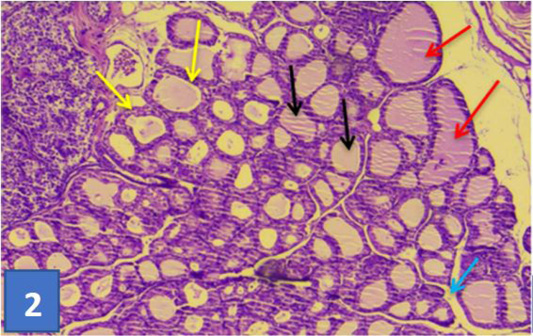
Figure 2:
Section of thyroid gland of control group: showing normal histological appearance of follicles (red darts) of different size lining with simple epithelial (yellow darts), colloid substance (black darts), this connective tissue septa separated gland lobules (blue dart). (H & E) stain (10X).