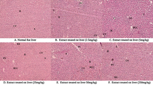
Figure 7:
Photomicrographs of liver sections following single oral administration of the ethanolic extract of Schinus terebinthifolius Raddi fruits showing a normal histological structure of the hepatocyte (H), the sinusoid (S), Centrilobular vein (CV), Kiernan’s spaces (KS), Hepatic cell cords (HCC), Portal vein (PV), Portal area (PA), and Binucleate hepatocyte (H and E-stained under 20× magnification power).