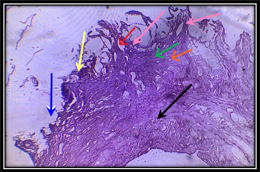
Figure 5:
A cross-section of the wall of the small intestine showing fibrosis in lamina muscularis mucosae( ), with the presence of blood bleeding ( ), inflammatory cells ( ), erosion ( ), degeneration of the mucosa layer( ) , with the appearance of ulcer( ) and irregularity of the muscular layer( ). (400X).
