
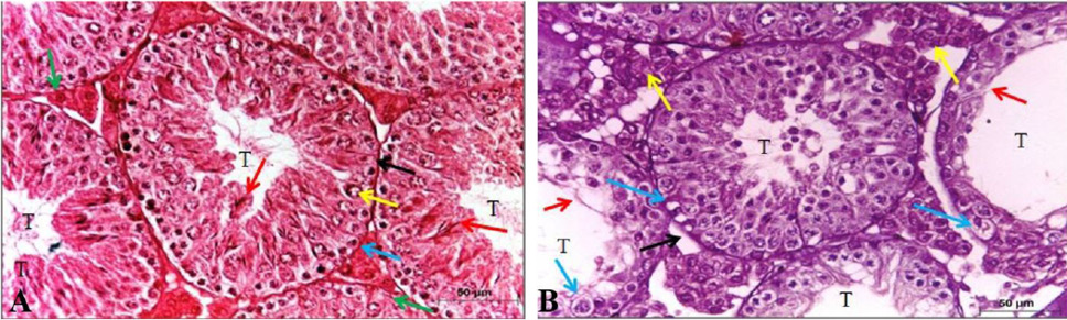
Fig. 1.
Effect of oral administration of exogenous testosterone on testes of albino rat. Histological structure of transverse section of testis of control male albino mouse showing A: average-sized seminiferous tubules (T) with average basement membranes (black arrow), spermatogonia (blue arrow), primary spermatocyte (yellow arrow), many sperms (red arrow) and average interstitium showing Leydig cells (green arrow). B: small-sized and dilated tubules (T) with thickened basement membrane (black arrow), thin germinal lining with scattered spermatogonia and primary spermatocytes with vacuolated cytoplasm (blue arrow), no sperms (red arrow), and excess interstitium with average Leydig cells (yellow arrow). Stain and Magnification H and E, X 400.