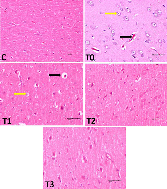
Figure 1:
Histological changes in the cerebral cortex of rats exposed to LA and treated with EBN. Note: The control group displayed a typical. The T0 group displayed haemorrhage and pyknotic nuclei, that considered signs of degenerative changes in neural cells. T1 and T2 groups demonstrated neuronal cell protection. T3 showed the regeneration, resemble in the histo-architecture for control.