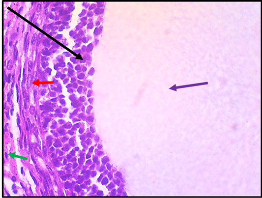
Figure 15:
Histological section in ovary of rat in (G2 group, CurSeNPs 10.47 μg/kg.bw.). The section shows graffian follicles (Black arrow), germinal epithelium (Red arrow), theca layer (Green arrows) and antrum (purple arrow). The tissue is stained with H and E stain and the section is captured using light microscope and digital camera at 40X magnifier scale.