
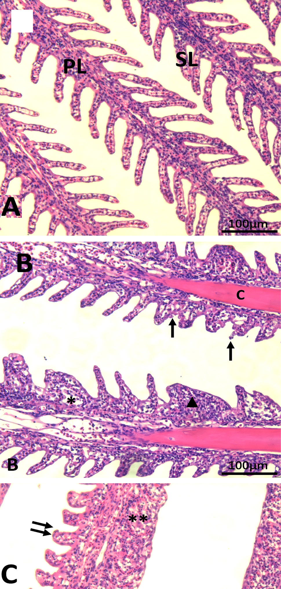
Fig. 1.
Transverse sections showing histopathology of gill(200X), A: Normal appearance (C group, 6 h); B: Hyperplasia in lamellar epithelium leading to lamellar fusion, epithelial lifting, curling of secondary lamellae; C: Hyperplasia in lamellar epithelium leading to lamellar fusion, epithelial lifting and occasional desquamation and disruption (T3 group, 96 h). Note: primary lamellae (PL), secondary lamellae (SL), epithelial hyperplasia (▲), epithelial necrosis and desquamation (↑), curling of secondary lamellae (↑↑), epithelial lifting and oedema (*), lamellar fusion (**).