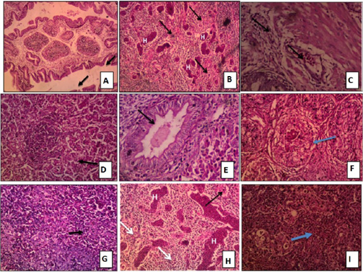
Figure 5:
Tissue sections stained with H and E of Chicken groups challenged with virulent NDV and IBDV showing:
A: Bursa: sever depletion of lymphoid follicles accompanied with inter follicular connective tissue deposition and hypertrophy of the mucous line forming finger like projections H and E x100. B: Bursa: sever depletion of lymphoid follicles with intra follicular hemorrhage H and E x100. C: Intestine: hemorrhage in sub mucosa with lymphocytic infiltration H and E x200. D: Liver: Hydropic degeneration in hepatocytes cytoplasm and focal area of coagulative necrosis infiltrated with lymphocytes H and E x 200. E: Liver: vacuolar degeneration in the cytoplasm of hepatocytes and hypertrophy of bile duct lining epithelium forming finger like projection in the lumen H and E x 200. F: Spleen: fibrosis of the lymphoid follicles H and E x200. G: Spleen: depletion of lymphoid follicles of white pulp H and E x 200. H: Thymus: focal area of hemorrhage, congestion of medulla, congestion of thymic artery and depletion of medulla H and E x 200. I: Thymus: depilation of cortex H and E x200.