
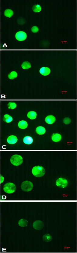
Fig. 2.
Fluorescent photomicrographs, 200× magnification. A, group of control; B, 2-cell; C, 4-cell; D, 8-cell; E, 16-cell.
Fluorescent photomicrographs, 200× magnification. A, group of control; B, 2-cell; C, 4-cell; D, 8-cell; E, 16-cell.