
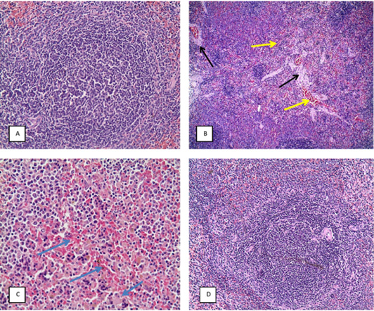
Figure 8:
(H&E X20), A: spleen of CG, B: spleen of TG showing splenic hemorrhage and congestion (yellow arrow), vascular and perivascular edema (black arrow) and depletion of white pulp, C: spleen of DAG showing splenic hemorrhage (blue arrow), edema and depletion of white pulp, D: spleen of BG showing more or less to normal.