
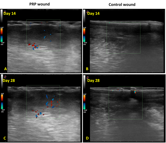
Fig. 1.
Ultrasonographical evaluation of neo-vascularization in the cutaneous wound of both groups. A; PRP treated wound obtained at day 14 showed mild development of blood vessels. B; Control wound showed no development of neo-vascularization at day 14. C; PRP treated wound at day 28 showed increased development of neo-vascularization at day 28. D; Control wound at day 28 showed mild development of neo-vascularization.