
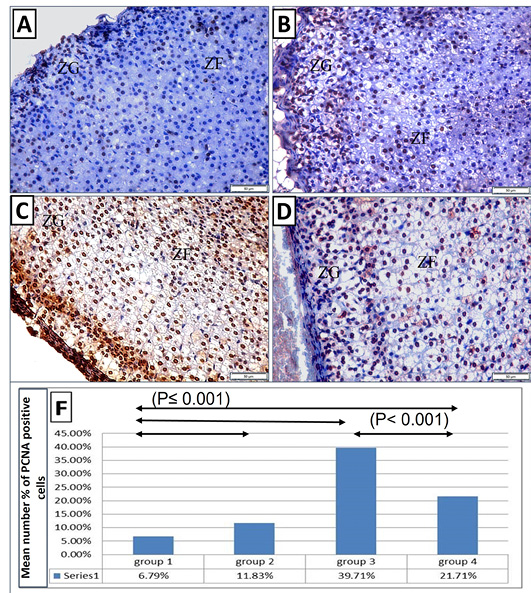
The immunohistochemical reaction of PCNA stained sections (X 400): (A) control group showing few sporadic immunoreactive PCNA positive cells with nuclear reaction in zona glomerulosa and zona fasciculata. (B) Control exercise group showing obvious increase of the PCNA positive cells in zona glomerulosa and zona fasciculata. (C) Depression group showing enormous increase of the PCNA positive cells with intense nuclear reaction in zona glomerulosa, at the junction between in zona glomerulosa and zona fasciculata and dispersed throughout zona fasciculata. (D) Depression exercise group showing less PCNA positive cells than those in the depression group, with the stained cells dispersed in zona glomerulosa and zona fasciculata. (E) The mean numbers % of PCNA positive cells in different groups.