Figure 4:
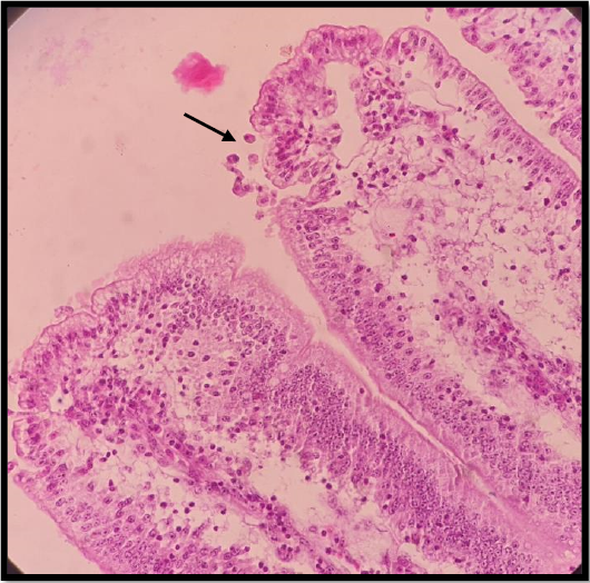
G2 (14 dpi), Cryptosporidium on the epithelial surface lining (oval round ball) (black arrow), with proprial distention tissue with leukocytic infiltration (H & E stain X 40).
G2 (14 dpi), Cryptosporidium on the epithelial surface lining (oval round ball) (black arrow), with proprial distention tissue with leukocytic infiltration (H & E stain X 40).