
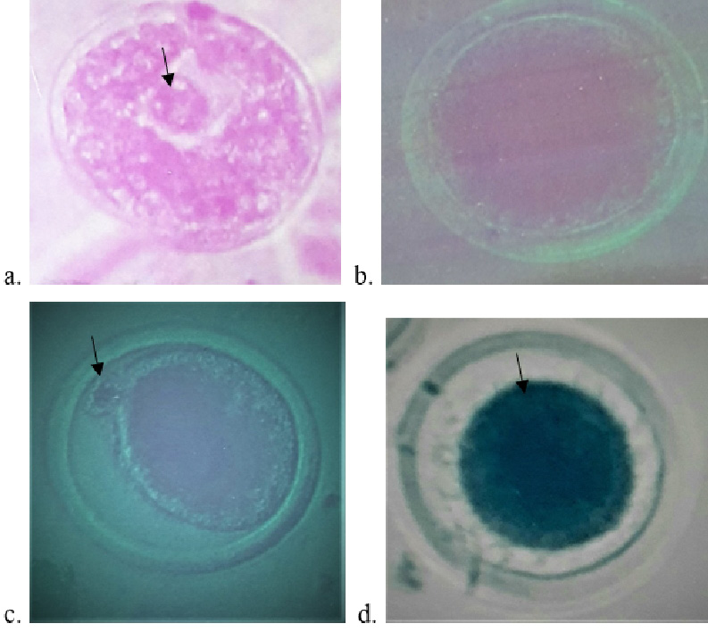
Fig. 1.
Photomicrographs of fully grown Nili Ravi buffalo oocytes during in vitro culture: (a) An intact germinal vesicle (indicated by arrow) with a single prominent nucleus arrested in dictyate stage of the first meiotic prophase. (b) Oocyte undergoing GVBD showing complete dissolution of the GV with only a small remnant of the nucleolus. (c) MII oocyte arrested at metaphase II stage and omitted a polar body (indicated by arrow). (d) Degenerated oocyte showing shrinkage of ooplasm. Photographs were taken from an inverted phase contrast (Nikon) microscope (objectives: a-d, X20).