
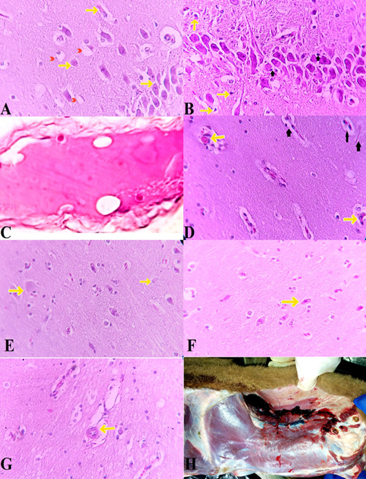
Brain changes in Traumatic Brain Injury. The early changes reflect alterations in ion gradients accompanying shifts in the perikaryon (Soma) and cause cloudy swelling. A: Swelling of the endoplasmic reticulum appears as a gap (cleft) in the periphery of the soma (Yellow arrows). This is different from crystalline inclusions and should not be mistaken. Astrocyte takes up Potassium + and the cytoplasm becomes pale due to intracellular fluid accumulation (red arrowhead) (H&E, x600). B: In response to brain-contusions injuries, neurons appear dark and pyknotic. (yellow arrows) (H&E, x200). astrocytes as well as Surveillant microglia activated to produce extra inflammatory mediators (Black Arrows). C: due to cellular edema and sufficient concentration of swollen neurons, some regions appear pale. (H&E, x12.5). D: necrotic neurons have a uniform eosinophilic cytoplasm. (yellow arrows). highe proportion of activated microglia and astrogliosis (Black Arrows) (H&E, x600). E: Some neurons become ghost cells without nucleus staining and a barely visible cell architecture (yellow arrows). (H&E, x600). F: rare eosinophilic neurons (yellow arrows) (H&E, x600). G: Toxoplasma gondii tissue cysts in the cerebral cortex (yellow arrows). (H&E, x600). H: Traumatic myositis in Cleidomastoid, Sternomastoid, Sternothyroid, Sternohyoid and Trapezius Muscles and sever damage to jugular vein, linguofacial vein, maxillary vein and caudal auricular vein, great auricular nerve lead to ruptures and hemorrhage.