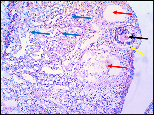
Figure 12:
Histopathological section in ovary of rat that treated with doxorubicin group G1. (4.4mg./kg.b.w. for 14 days). The section shows clear congestion of graffian follicles (Oocyte, Black arrow) with sloughing and degenerative lesion in the granulosa cell layer (Yellow arrow). The section shows clear necrotic lesion in the graffian follicle with lysis of Oocyte (Red arrows). The Oogonium shows clear damage and necrosis of epithelia (Blue arrows). The tissue is stained with H&E stain and the section is captured using light microscope and digital camera at 10X magnifier scale.