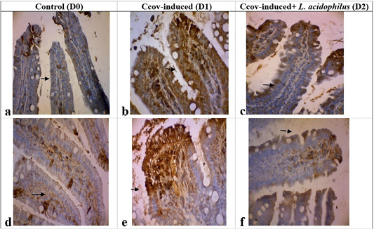
Figure 3:
Positive TNF-α expression, shown as brown aggregates in the intestinal mucosa and submucosa duodenal (b), was significantly higher compared to the non-reactive control group D0 (a). in contrast, TNF-α expression in the isntestinal villi showed a moderate decrese (c) with non-specific IL-1β expression in the epithelia and intestinal crypts (d). Strong IL-1β immunoreaction, depicted as brown aggregates in the intestinal villi’s epithelial and lamina propria area (e), contrasted with the weak IL-1β immunoreaction in the lamina propria area (immunohistochemistry, hematoxylin counterstained, arrows indicate morphological changes).