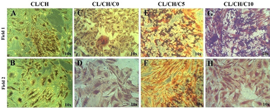
Figure 8:
von Kossa staining of osteoblastic cells of first passage/ third week at day 21 cultured in osteogenic medium barrel to cells were seeded with the various of scaffolds extract (A-H) two fields for each scaffold extract brown -black mineral deposits (Mineralized areas) were seen by large cell colony in different fields of culture well, Inverted Phase Contrast Microscope Digital Camera Olympus, Magnification 10x.