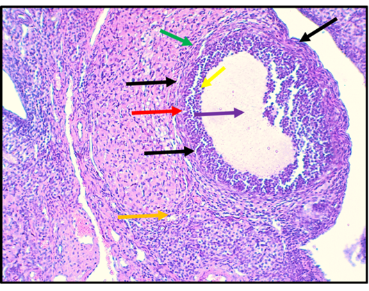
Figure 14:
Histological section in ovary of rat in G2 group (CurSeNPs 10.47 μg/kg.bw.). The section shows graffian follicles (Black arrows), germinal epithelium (Red arrows), theca layer (Green arrows), granulosa cell layer (Yellow arrows), antrum (purple arrow) and Oogonium (Orange arrow). The tissue is stained with H and E stain and the section is captured using light microscope and digital camera at 10X magnifier scale.