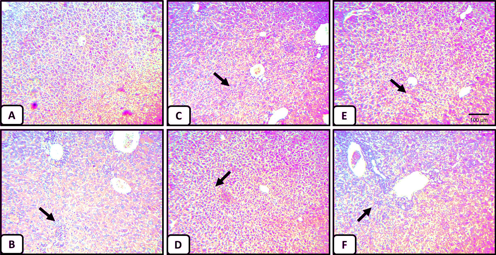
Histopathology of liver tissues. (A) Liver section of normal negative control mice (vehicle) shows central vein surrounded by hepatic cord of cells (normal architecture), (B) liver section of CCl4-treated mice showing massive fatty changes, focal central vein congestion, a variety of cavitations and necrosis in hepatocytes with inflammation, and loss of cellular boundaries (indicated by arrow), (C) liver section of mice treated with 100mg/kg showing normal liver architecture (magnification 5X), (D) liver section of mice treated with the 500mg/kg showing normal liver architecture, (E) liver section of mice treated with CCl4 and 100mg/kg of M. forsskalii showing absence of cavitations, necrosis, and inflammatory cells, and regeneration of hepatocytes around central vein toward near normal liver architecture but slight congestion in central vein (indicated by arrow), and (F) liver section of mice treated with CCl4 and 500 mg/kg of M. forsskalii showing mild central vein congestion (indicated by arrow), ballooning, and necrosis with sinusoidal dilatation H and E x 50.