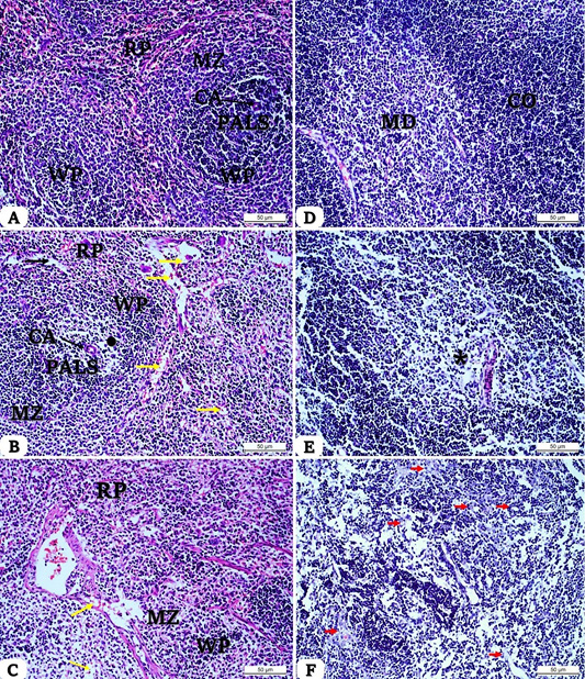
Histomorphology of control spleen (A) showed a normal appearance of red pulp (RP), white pulp (WP) with a well-differentiated marginal zone (MZ), central artery (CA), and periarteriolar lymphoid sheath (PALS). (B) The section of the spleen from the low-dose-dietary soy (6.6%) phytoestrogens group showed a mild depletion of T cells in PALS. Signs of lipidosis were also detected (yellow arrows). (C) Severe lymphocytes depletion in the WP compartments and RP was observed with high-dose dietary soy (26.41%) phytoestrogens in the spleen section (*). (D) The thymus (control group) displayed normal architecture of the cortex (CO) and medulla (MD). (E) Thymus section of a low-dose-dietary soy (6.6%) phytoestrogens group showed a cellularity decrease in the medullary area (*). (F) Thymus section of a high-dose-dietary soy (26.41%) phytoestrogens group exhibited severe depletion of lymphocytes in the cortex, in addition to the presence of apoptotic lymphocytes (red arrows), [H&E stain; original magnification: ×20].
