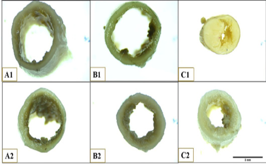
Figure 2:
Photograph illustrates the lumen and wall of Muscovy duck’s caecum at anterior end of proximal portion (A1), posterior end of proximal portion (A2), anterior end of middle portion (B1), posterior end of middle portion (B2), anterior end of distal portion (C1), posterior end of distal portion. Scale bar =3mm