
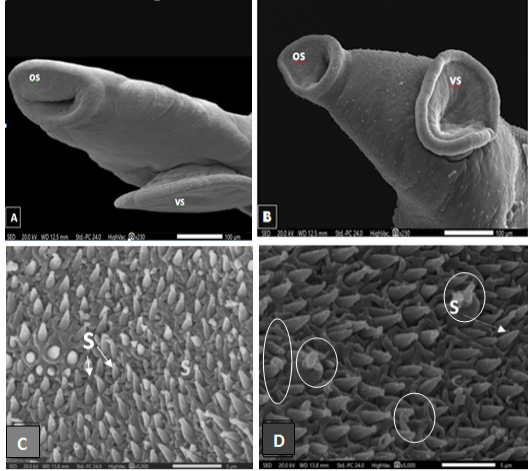
Figure 2:
Scanning electron micrographs of S. mansoni worm collected from (A) Infected control mouse showing an anterior end with normal oral (os) and ventral suckers (vs). (B) Infected mouse treated with FWE showing no clear change in the oral and ventral suckers. (C) Enlarged portion of (A) showing oral sucker region covered with sharp spines. (D) Enlarged portion of (B) showing some corrosions in oral sucker spines after FEW treatments (circled).