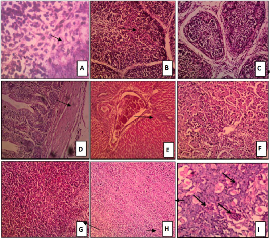
Figure 3:
Tissue sections stained with H and E of chicken groups received challenged by NDV only.
A: Bursa: activation of germinal center that showing mitotic figure H and E x 400. B: Bursa: depletion of lymphoid follicles H and E x 200. C: Bursa: inter follicular edema and severe depletion of lymphoid follicles. D: Intestine: mild lymphocytic infiltration of the mucosa H and E x200. E: Liver congestion of central vein H and E x100. F: Liver: focal area of liver necrosis characterized by lymphocytic infiltration H and E x 200. G: Spleen: congestion of red pulp H and E x 200. H: Spleen; sever depletion of lymphoid follicles H and E x100. I: Thymus sowed hemorrhages and necrosis of medulla H and E x 200.