Figure 1
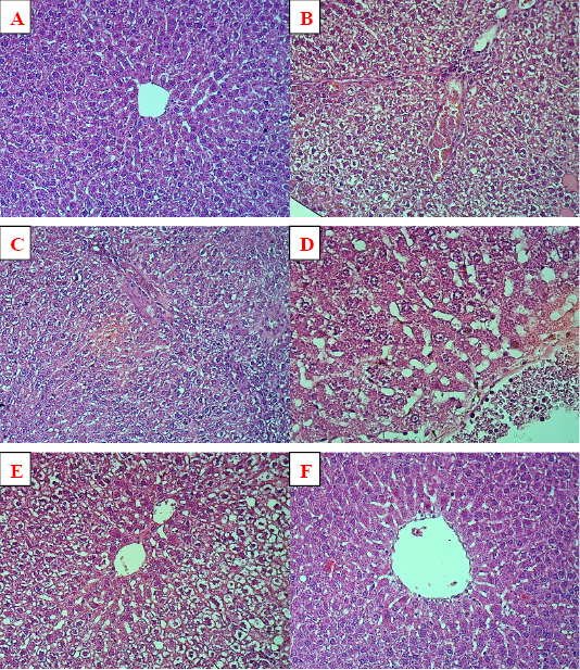
Histopathology of liver tissue. (A) negative control with normal hepatic tissue architecture, X20. (B) positive control showed fatty changes of hepatocytes along with congestion of blood vessels and infiltration of inflammatory cells, X20. (C) and (D) rousovastatin groups showed vacuolation of hepatocyte with fatty changes and congestion of blood vessels with inflammatory cells, X20, X40 respectively. (E) combination group showed accumulation of fat droplets inside the hepatocytes, X20. (F): pumpkin group presented with mild degree of fatty changes and appear normal in other one to some degree with normal architecture, X20.
