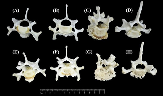
Figure 4
Spondylosis deformans lesion characterized by the osteophyte formation developed at the edge of the ventral vertebral body of C6 (A and E), C7 (B and F), T1 (C and G) and T12 (D and H). Bone degradation, characterized by erosion, roughened and irregular bone texture, was found on both the cranial and caudal intervertebral surfaces of all the affected vertebrae. (A-D) Cranial view; (E-H) Caudal view.