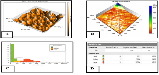
Figure 7:
Atomic force microscopy (AFM) was used to analyze the size distribution of selenium nanoparticles (CurSeNPs). The results were shown in the form of a two-dimensional picture (a), a three-dimensional image (b), and a size distribution histogram (c) and (d).