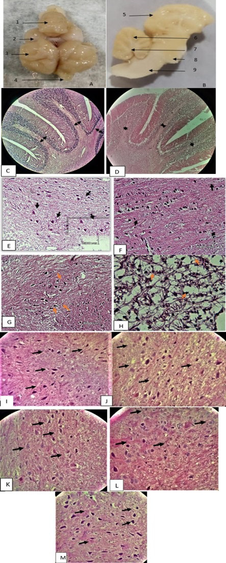
Cerebellar and cerebral white matter (H & E x 20 and x 40). (A) positive control entire brain (1: cerebellum; 2: optic lobe; 3: cerebral hemisphere; 4: olfactory lobe). (B) positive control sagittal brain section (5: cerebral cortex; 6: cerebellar cortex; 7: deep cerebellar white matter; 8: pons; 9: medullar oblongata. (C) positive control after six months shows normal thickness of cerebellar white matter folds. (D) six months CPZ treated animals show loss of cerebellar thickness in white matter folds. (E) control group kept for six months without CPZ shows deep cerebellar white matter and normal oligodendrocyte population. (F) animals with CPZ treatment for six months show rare oligodendrocytes. (G) cerebral white matter of control animal after six months shows normal glial cells. (H) cerebral white matter of six months CPZ treated animal shows apoptotic oligodendrocytes (arrow) and shrinkage of axons. (I) normal oligodendrocyte population can be seen in control animal after seven and half months without any treatment. (J) spontaneous oligodendrocyte regeneration can be seen in animal with no treatment for six weeks after six months of CPZ treatment. (K) honey treated group showing regeneration of oligodendrocytes. (L) black seed treated group showing increase in glial cells population. (M) Mixture (honey and black seed) treated group shows significantly increased oligodendrocyte population.