
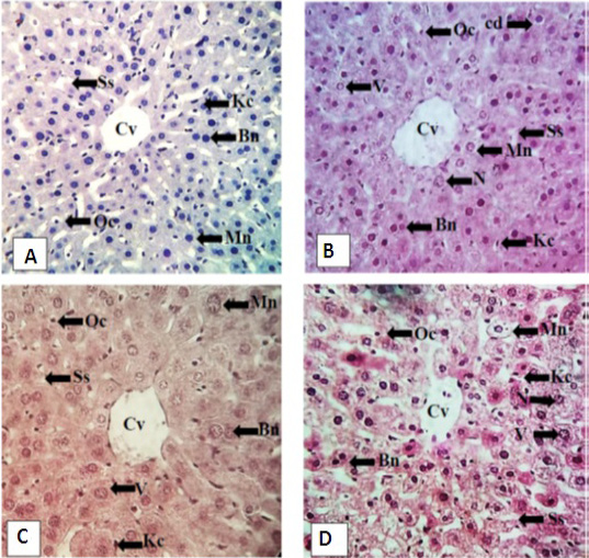
Effect of exposure of feed to microwaves in different contains on histological structure of liver of male mice. A: Control showing well organized mononucleated (mn) and bi nucleated (bn) hepatocytes of similar size in cord like structure having round nuclei. Sinusoidal spaces (Ss) of equal width lies between hepatic cords, round oval cells (Oc) observed in sinusoidal spaces. Central vein (Cv) is compact. Kupffer cells (Kc) elongated in shape lining the hepatocytes. B: Direct group represented decreased number of hepatocytes as well as nuclear vacuolation (V), necrosis (N) and cellular degeneration (cd). C: Glass group showed decreased number of hepatocytes (Bn and Mn) of variable size and shape, nuclear vacuolation and necrosis. Dilated central vein with infiltrated cells in its lumen. Sinusoidal spaces became wider. D: Plastic group also showed dilated sinusoidal spaces and central vein having infiltrated cells. Number of oval and Kupffer cells was decreased and nuclear vacuolation was also observed. Stain: hematoxylin and Eosin Magnification: 400X.