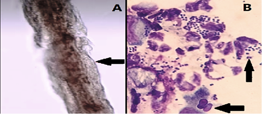
Figure 2
A: Infected hair with Microsporium. Note the swollen distorted appearance of the hair shaft with masses of ectothrix spores clustered around it, (X 40). Infected hair appear swollen, frayed, irregular and fuzzy in outline, and the normal structure of cuticle, cortex, and medulla is lost, beaded chains of small rounded cells (spores) and hyphae seen uniform in diameter (arrow), septate and variable in length and degree of branching.
B: Ear cytology sample from a cat patient with Otitis externa. Numerous Streptococci are present in pairs (upper arrow), also pictured are numerous inflammatory cells exhibiting bacteriophagy (lower arrow) and an epithelial cell. (X100, Romanowsky-type stains).
