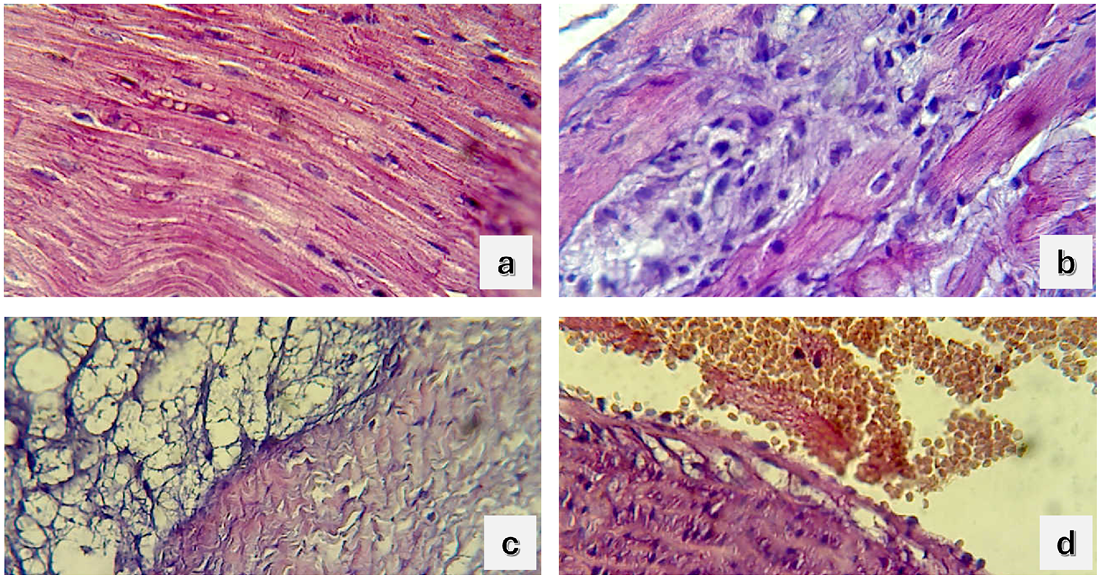
Figure 2A:
Section in normal cardiac muscle architecture in control group (G1); 2B: Section in the heart of group 2 shows no mononuclear cells infiltration between cardiac muscle; 2C: Section in the heart of group 2 shows necrotic cardiac muscle with vacuole of fatty droplets; 2D: Section in the aorta of G4 group shows vacuolation in subintima (H andE stain 400X).