
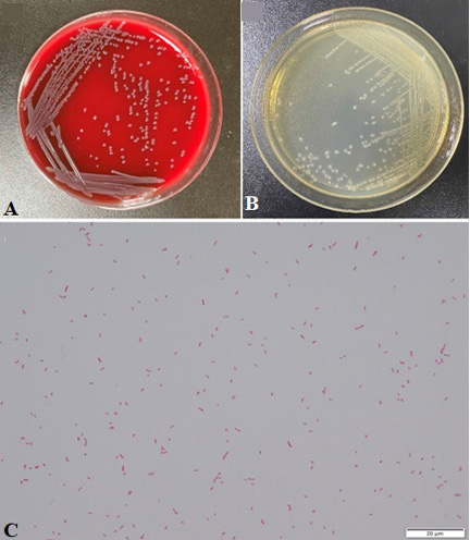
Fig. 2.
Morphologies of C. freundii FSN1. A: Colony morphologies of FSN1 on LBA plate; B: Colony morphologies of FSN1 on 5% sheep blood agar; C: Gram staining of bacterium FSN1 (100 ×). Scale bars = 20 μm.
Morphologies of C. freundii FSN1. A: Colony morphologies of FSN1 on LBA plate; B: Colony morphologies of FSN1 on 5% sheep blood agar; C: Gram staining of bacterium FSN1 (100 ×). Scale bars = 20 μm.