Figure 10:
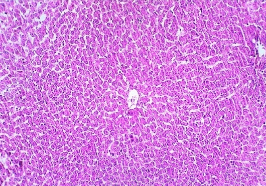
Normal histological structure of liver in control group mice. The prepared section was stained with Hematoxylin and Eosin stain and examined at 10X. The image is represented experimental mice.
Normal histological structure of liver in control group mice. The prepared section was stained with Hematoxylin and Eosin stain and examined at 10X. The image is represented experimental mice.