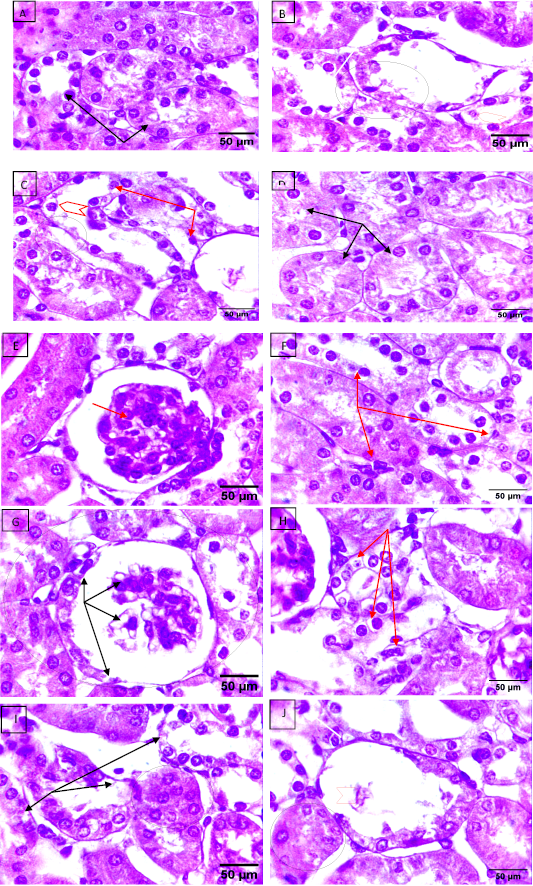
Figure 1:
Light micrographs of the rat’s kidney sections, (stained with H and E. magnification power 1000X). Group I- A and Group 3- D and E show normal proximal and distal convoluted tubule structures (black arrows) and glomerulus structures (red arrows). Groups 2 B and C, show severe renal injury in the proximal and distal tubules, represented by lumen dilation (black arrows), vacuolization of tubule lining cells (red arrows head), degeneration of some tubules cells (red arrows), and necrosis (circle). Groups 4- F and G show much improvement in the kidney glomerulus and renal tubule structure (circle), except some histological damage was still seen in the proximal tubules (red arrow) and renal corpuscle (black arrows). Group 5- H, I, and J, shows the renal injury was shown loss of glomerulus (arrowhead), renal tubular necrosis (black arrows), cytoplasmic vacuolation (red arrows), and hypertrophy of proximal convoluted lining cells and close of tubules lumen (circle).