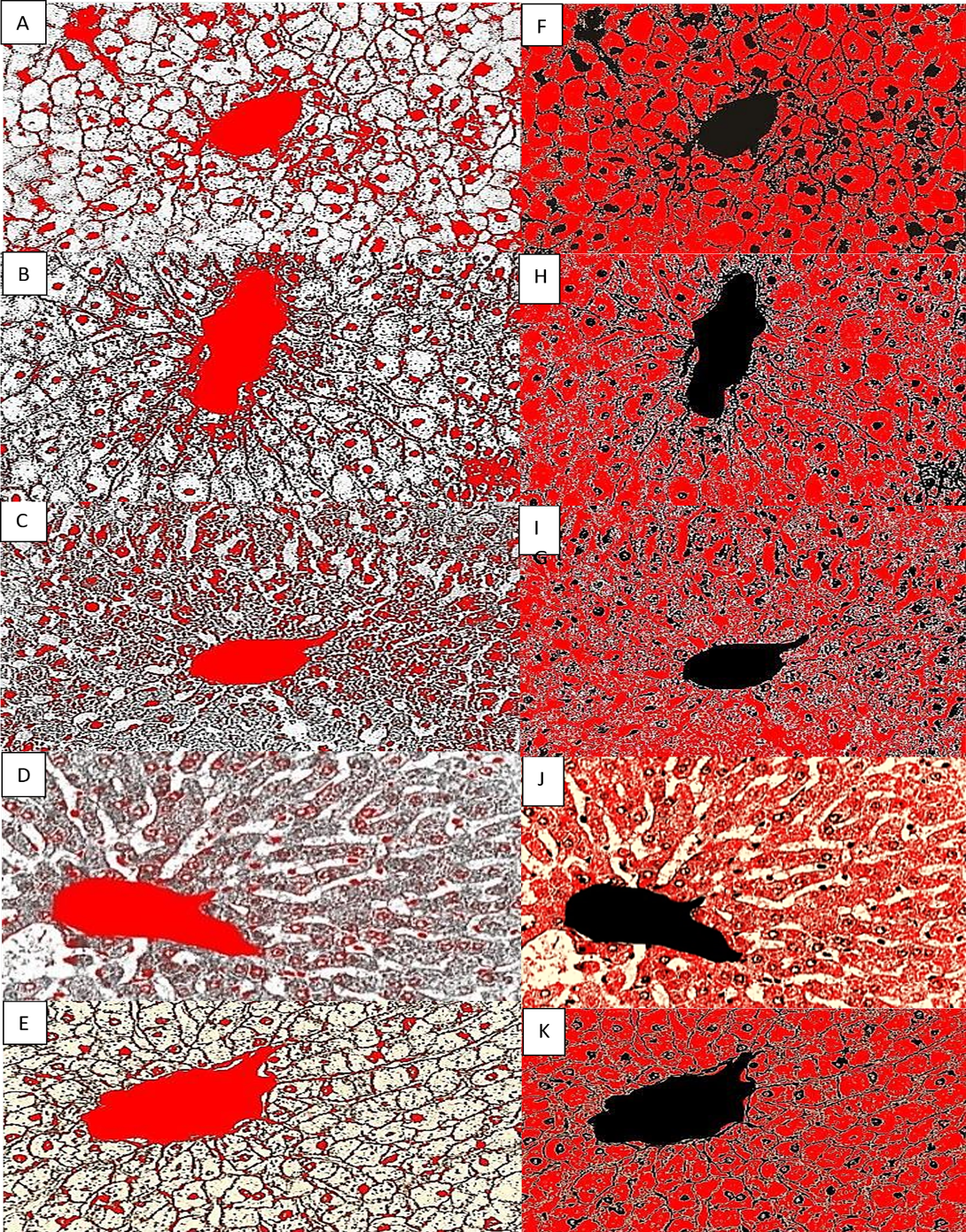
Figure 3.
Photomicrographs (400X) of liver tissue specimens were image-analyzed using Image J software as a percentage of quantification for nuclei and cell cytoplasm (a, b, c, d, e, and f). The samples were fixed in formaldehyde for 12, 24, and 48 hours. While (g, h, I, j, k, and l) were fixed in Bouin’s fluid for 12, 24, and 48 hours, and (m, n, p, and q) were fixed in Carony,s fluid for 12 and 24 hours.