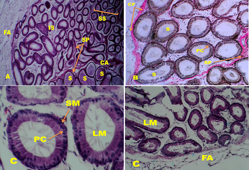
Microscopic section of epididymis segments (A) Initial (IS) and caput segments (CA) of Wistar Rats aged 66 days in non-treated groups showed were surrounded by friable amount of fat (FA) and divided into sub-segmental division (SS), by loose connective tissue septa (SP) rich with collagen fibers (CF). Principle cells (PC) one type of epithelial cells lined tubules that full with sperms. (A) HandE 40X; (B) Initial (IS), of non-treated groups (G1) showed ducts surrounded by clear quantity of collagen fiber(CF) and ducts divided by loose connective tissue septa (SP), each ducts full with sperm(S)Van Gieson’s stain 100X(C) Initial segments of treated groups(G2) showed lumen duct (LM) devoid of sperm, each tubule surrounded from out by layers of smooth muscle (SM), and lined by epithelia that contain most cell such as principle cell(PC). (C)HandE 400X (left picture), 100X (right picture).