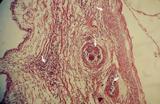
Figure 7:
The micrograph of the intestine shows the orientation of cellular organization disintegrated by fibrotic infiltration. Eosinophils are scattered throughout the section due to heavy infection of cestodes and nematodes (Ascaridia sp. and Capillaria sp.) (Arrow) × 200.
