
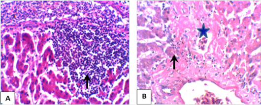
Figure 1:
Representative photomicrograph of gander livers in the control group showing: (A) extensive perivascular leukocytes aggregations infiltrating the hepatic parenchyma (arrow), and (B) marked perivascular hepatic necrosis (star) and lymphocytes admixed with few macrophages and fibroblasts (arrow). (H & E, x400).