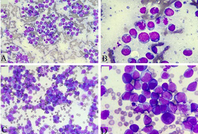
Fig. 1.
Microscopic pictures showing the typical cytological appearance of a high-grade lymphoma in lymph nodes and pleural effusion test (A and C, 40×; B and D, 100×). The smear of lymph node FNA showed the abundant cells especially lymphocytoblast.