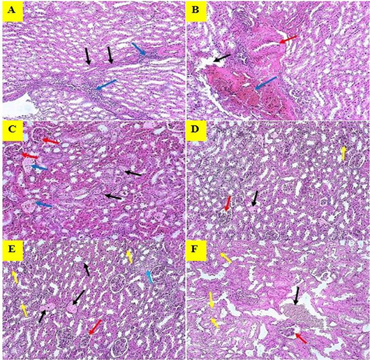
Histopathological changes of kidney in mice received estrogen. (A): Damaging of proximal tubules (Black arrow) and Inflammatory cells infiltration (Blue arrow); (B): Damaging of venous wall (Black arrow), Venous congestion (Blue arrow), Narrowing of ureter’s lumen (Red arrow); (C): Amyloid degeneration (Black arrow), Amyloid infiltration in veins (Blue arrow), Amyloid infiltration in proximal tubules (Red arrow); (D): Congestion of veins (Black arrow), Atrophy of glomerular tufts (Red arrow), Hypertrophy of proximal tubules (Yellow arrow); (E): Congestion of veins (Black arrow), Parenchymal fibrosis (Blue arrow), Atrophy of glomerular tufts (Red arrow), Hypertrophy of tubular lumen (Yellow arrow); (F): Congestion of veins (Black arrow), Hypertrophy of glomerular space (Red arrow), Hypertrophy of tubular lumen (Yellow arrow). The sections were stained with Hematoxylin and Eosin stain, examined at 10X, and the images are represented experimental mice.