
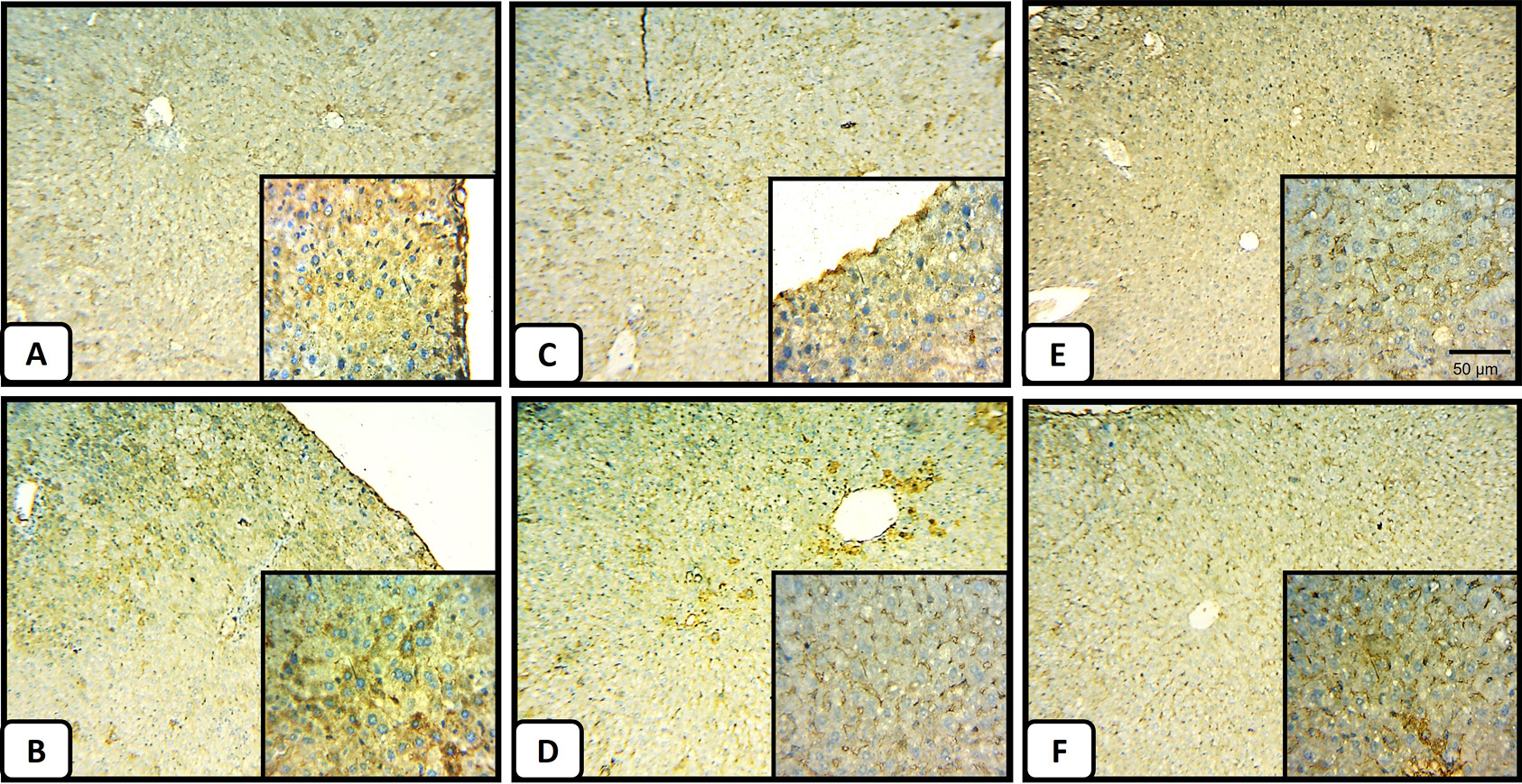
Immunohistochemistry of P53 in the liver tissues. (A) Section from a negative control mice liver shows the normal pattern of P53 with mild staining. (B) The liver section obtained from CCl4-intoxicated mice shows extensive staining (indicated by brown color) for P53. (C) and (D) a liver section from normal mice treated at low and high doses, respectively, showing a non-appreciated difference in P53 staining pattern (magnification, x 50 for the main photo and x 100 for the hyper-focused region in the small box at the left bottom of each panel). (E) and (F) Liver tissue sections prepared from the 100 mg/kg and 500mg/kg of M. forsskalii after CCl4 treatment, respectively, show less P53 staining compared to (B), however (E) shows a closer staining pattern to the normal liver (A).