

Quercetin Modulates Aging Induced-Testicular Damage Via Increasing the Antioxidant Defense and the Immunolocalization of the Proliferating Cell Nuclear Antigen in Male Rats


Quercetin Modulates Aging Induced-Testicular Damage Via Increasing the Antioxidant Defense and the Immunolocalization of the Proliferating Cell Nuclear Antigen in Male Rats
Eman A. Al-Shahari1,2, Abdelhalim A. Alkhazendar3, Eman R. ElBealy4 and Abeer A. Alm-Eldeen3*

Effect of quercetin on the levels of testes SOD (unit/ mg protein), MDA (nmol/ mg protein) and 8-OHdG (ng/ L) in 3 and 30 months old rats before and after quercetin administration. Data are means ± SD with n=4, Different letters indicate significant differences among the columns (p ≤ 0.05).
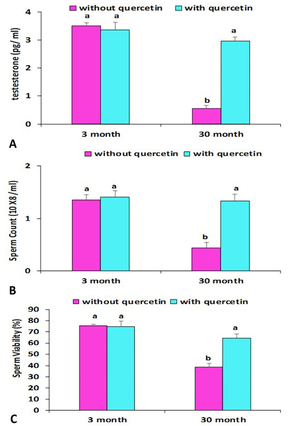
Effect of quercetin on the testesterone (pg/ ml), sperm count (10 X8 /ml) and sperm viability (%) in the 3 and 30 months old rats before and after quercetin administration. Data are means ± SD with n=4, Different letters indicate significant differences among the columns (p ≤ 0.05).

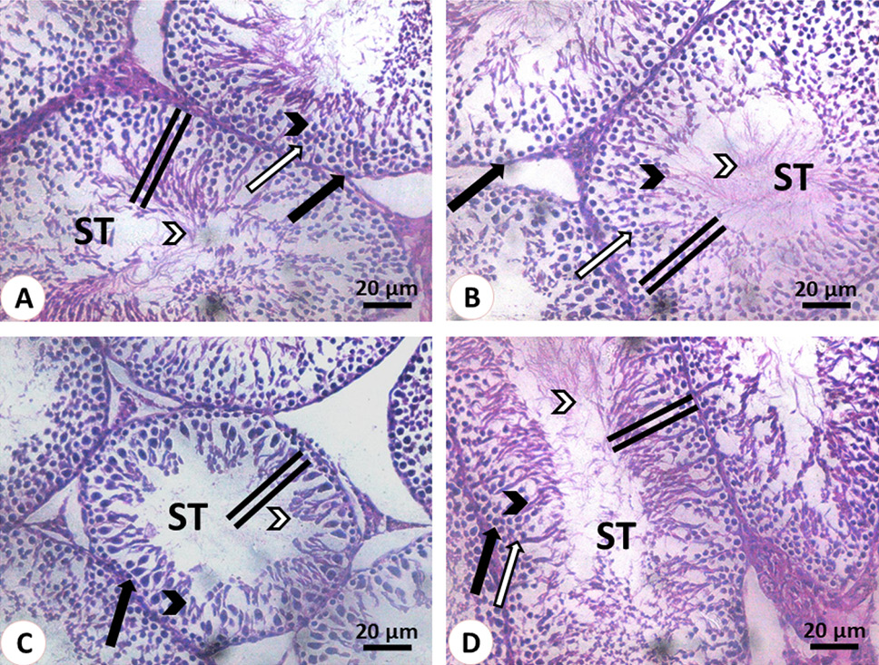
Histological structure of the testis stained with hematoxylin and eosin. (A, B): 3 months old rats before and after quercetin administration, respectively. The seminiferous tubules (ST) showed normal seminiferous epithelium (double line) that contains spermatogonia (black arrow), spermatocytes (white arrow), spermatids (black arrowhead) and mature sperm (white arrowhead). (C, D): The seminiferous tubules (ST) of 30 months old rats before quercetin administration in C showed the depletion of spermatogenic cells (black arrow), round spermatids (black arrowhead) and sperms (white arrowhead). Note the reduction in the seminiferous tubules diameter and its epithelium height (double line). The seminiferous tubules (ST) of 30 months old rats after quercetin administration in D restore the normal structure. Note the height of its epithelium (line), spermatogonia (black arrow), spermatocytes (white arrow), spermatids (black arrowhead) and mature sperm (white arrowhead).

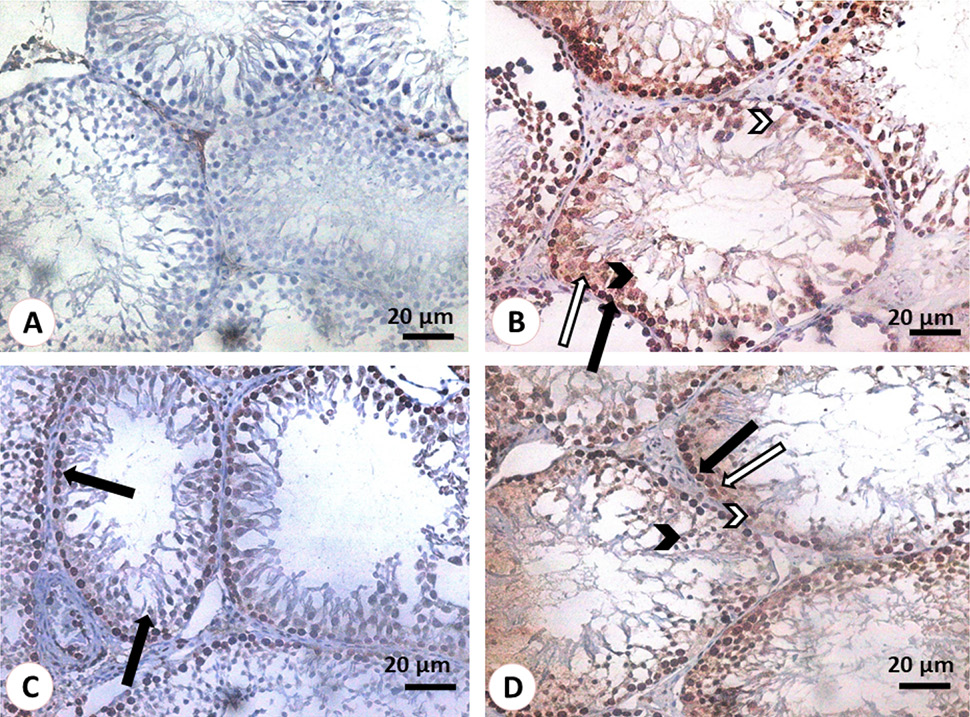
Histological structure of the testis stained PCNA. (A, B): 3 months old rats before and after quercetin administration, respectively. In A, no reaction was observed was used as as we omitted PCNA antibody from the reaction and used it as a negative control. In B, Spermatogonia (black arrow), spermatocytes (white arrow), spermatids (black arrowhead) and Sertoli (white arrowhead) were positively immunostained with PCNA. (C, D): 30 months old rats before and after quercetin administration, respectively. In C, only spermatogonia (black arrow) were positively immunostained with PCNA. In D, spermatogonia (black arrow), spermatocytes (white arrow), spermatids (black arrow head) and Sertoli (white arrowhead) were positively immunostained with PCNA.


