

Biochemical and Histopathological Effects of Repeated Low Oral Doses of Malathion, Metalaxyl and Cymoxanil on Different Tissues of Rats


Biochemical and Histopathological Effects of Repeated Low Oral Doses of Malathion, Metalaxyl and Cymoxanil on Different Tissues of Rats
Ahmed H. Massoud1, Mohamed S. Ahmed2, Moustafa Saad-Allah1, Aly S. Derbalah1, Ashraf Albrakati3* and Ehab Kotb Elmahallawy4*
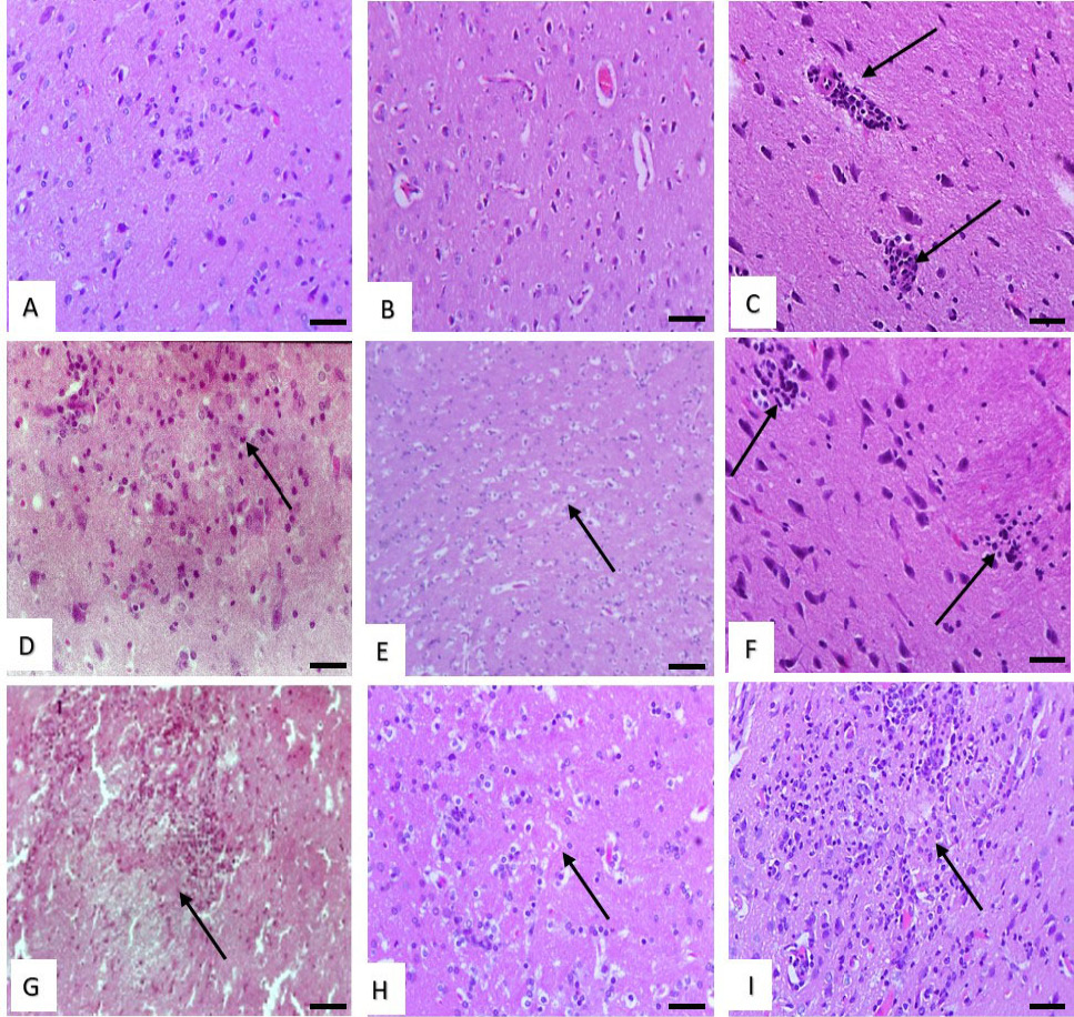
Histological structure of rat brain. (A and B) Effect of low dose of malathion and metalaxyl showing normal structure. (C) Effect of low dose of cymoxanil showing slight perivascular lymphocytes infiltration (arrows). (D) Effect of with medium dose of malathion showing neuronal cell degeneration (arrow). (E) Effect of medium dose of metalaxyl showing slight spongiosis (arrow). (F) Effect of medium dose of cymoxanil showing focal collections of glial cells forming Babe’s bodies (arrows). (G) Effect of high dose of malathion showing focal cerebral malacia (arrow). (H) Effect of high dose of metalaxyl showing cerebral spongiosis and gliosis (arrow). (I) Effect of high dose of cymoxanil showing cerebral liquifactive necrosis (malacia) (arrow). Stain: H and E. Magnification bar: 50 µm.

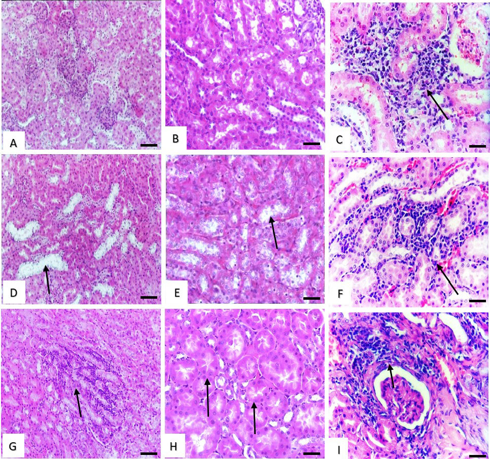
Histological structure of rat kidney. (A and B) Effect of low dose of malathion and metalaxyl showing normal structure. (C) Effect of low dose of cymoxanil showing, slight interstitial mononuclear cells infiltration (arrow). (D and E) Effect of medium dose of malathion and metalaxyl showing slight vacuolar degenerative changes in the renal tubules lining epithelium (arrows). (F) Effect of medium dose of cymoxanil showing slight interstitial nephritis (arrow). (G) Effect of high dose of malathion showing marked interstitial nephritis (arrow). (H) Effect of high dose of metalaxyl showing cloudy swelling in the epithelial lining of the renal tubules (arrows). Stain: H and E. Magnification bar: 50 µm.

Histological structure of rat lung. (A and B) Effect of low dose of malathion and metalaxyl showing normal structure. (C) Effect of low dose of cymoxanil showing slight interstitial mononuclear cells infiltration (arrow). (D) Effect of medium dose of malathion showing marked thickening in the interalveolar septa (arrow). (E) Effect of medium dose of metalaxyl showing slight interstitial pneumonia (arrow). (F) Effect of high dose of malathion showing more marked interstitial nephritis (arrow). (G) Effect of high dose of malathion showing, massive peribronchial infiltration of mononuclear cells (arrow). (H) Effect of with high dose of metalaxyl showing slight interstitial pneumonia (arrow). (I) Effect of high dose of cymoxanil showing marked interstitial mononuclear cells infiltration (triangle) and pneumocyte type II hyperplasia (arrows). Stain: H and E. Magnification bar: 50 µm.

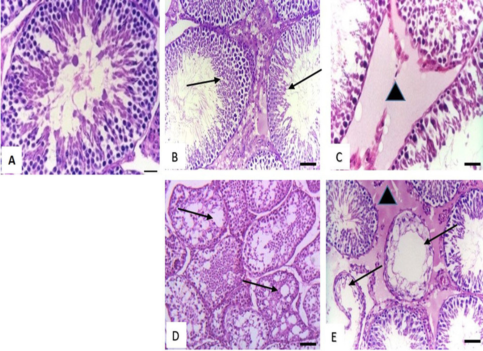
Histological structure of rat testis. (A) Effect of low dose of malathion, (low, medium, high) dose of metalaxyl and low dose of cymoxanil showing normal structure. (B) Effect of medium dose of malathion showing decrease in the number of spermatogenic cells in the seminiferous tubules (arrows). (C) Effect of medium dose of cymoxanil showing interstitial oedema (triangle). (D) Effect of high dose of malathion showing necrosis of spermatogenic layers and marked vacuolation in the cytoplasm of sertoli cells (arrows). (E) Effect of high dose of cymoxanil showing, massive necrosis in the lining epithelium of seminiferous tubules (arrows) with marked interstitial oedema (triangle). Stain: H and E. Magnification bar: 50 µm.

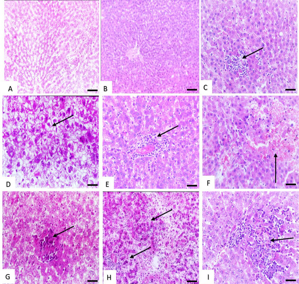
Histological structure of rat liver (A and B) Effect of low dose of malathion and metalaxyl showing, slight congestion and dilation of the hepatic sinusoids. (C) Effect of low dose of cymoxanil showing slight sinusoidal cell activation with pregranuloma formation (arrow). (D) Effect of medium dose of malathion showing marked cytosolic hydrops with vacuolated cytoplasm of the hepatocytes (arrow). (E) Effect of medium dose of metalaxyl showing perivascular focal collection of mononuclear cells (arrow). (F) Effect of medium dose of cymoxanil showing haemorrhages in the hepatic parenchyma (arrow). (G and H) Effect of high dose of malathion, metalaxyl and cymoxanil showing hepatocellular necrosis (arrows). Stain: H and E. Magnification bar: 50 µm.


