

Anatomical and Pathological Evaluation of Congenital Anomalies in Calves: 6 Cases


Anatomical and Pathological Evaluation of Congenital Anomalies in Calves: 6 Cases
Gülseren Kırbas Dogan1,*, Emin Karakurt2, Mushap Kuru3 and Hilmi Nuhoglu2
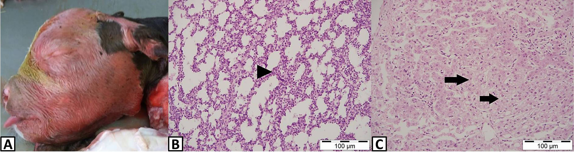
A, Anasarca. B, Lung, thickening of the alveolar wall (arrowhead), H&E, 100 μm. C, Liver, hydropic degeneration of hepatocytes (arrows), H&E, 100 μm.

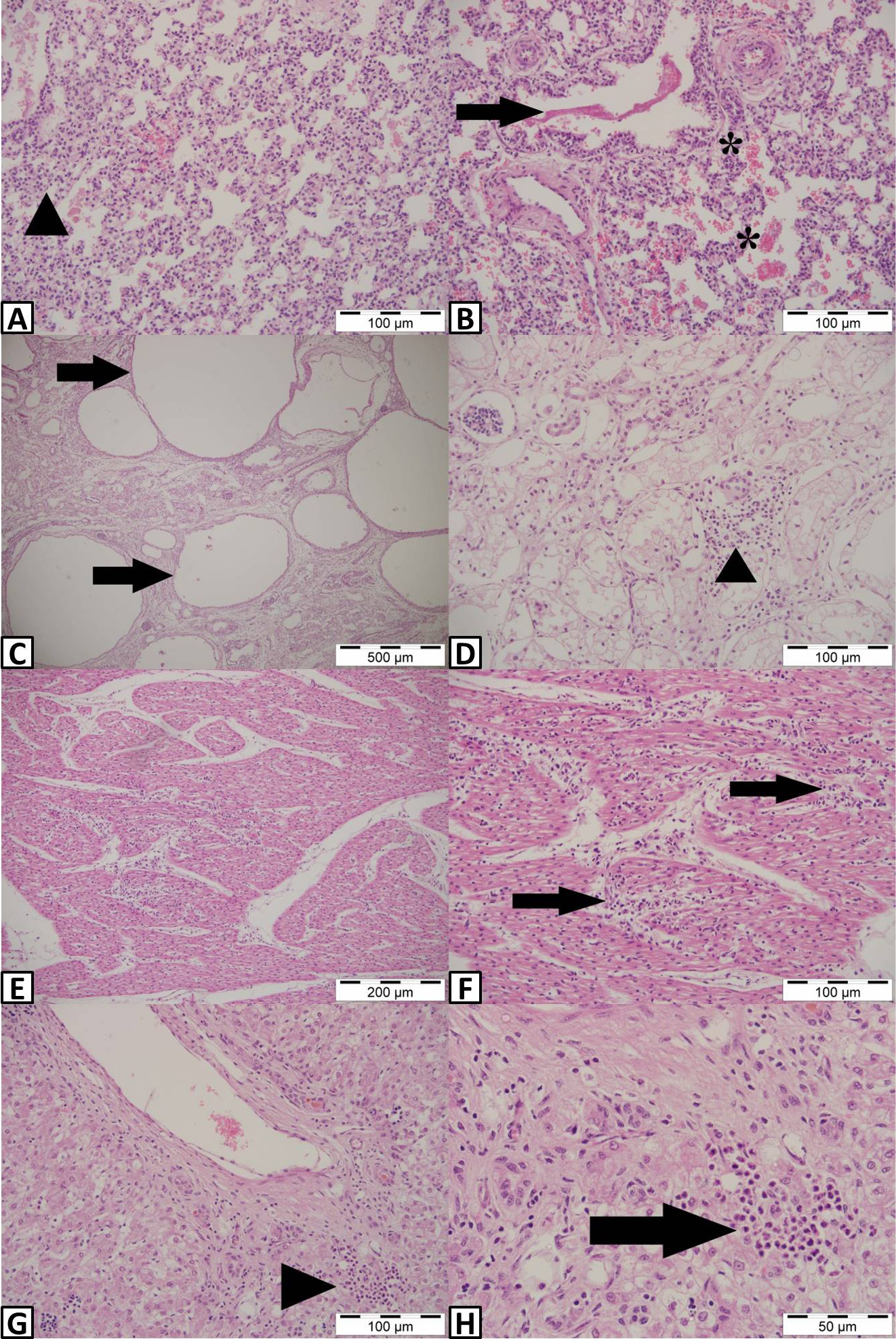
A, Lung, Atelectasis (arrowhead), H&E, 100 μm. B, Lung, Severe bleeding and hyperemia (stars), content in bronchioles (arrow), H&E, 100 μm. C, Kidney, cystic structures (arrows), H&E, 500 μm. D, Kidney, nonpurulent interstitial nephritis (arrowhead), H&E, 100 μm. E, Heart, nonpurulent myocarditis (arrows), H&E, 200 μm. F, Higher magnification, H&E, 100 μm. G, Liver, neutrophilic inflammatory cell infiltration (arrowhead and arrow), H&E, 100 μm. H, Higher magnification, H&E, 50 μm.

A and B, Two-nosed calf. C, Opening in the cranium. D, Brain, purulent meningitis (M), H&E, 500 μm. E, Higher magnification, purulent meningitis (arrowheads), H&E, 200 μm. F, Brain, vascular hyperemia (h) and inflammatory cell infiltration (arrow), H&E, 50 μm. G and H, Substansia grizea with meninges in the brain. H&E, 1000 μm. I, Synaptophysin positive areas in the region where the substantia alba in the area where substansia grizea is located in the inner brain region (arrows), IHC, 1000 μm. J, Synaptopysin positive areas in substansia grizea (arrowheads) and Synaptopysin negative areas in substansia alba, IHC, 500 μm. K, Brain, calcification (arrows), H&E, 50 μm. L, Brain, gliosis (arrowhead), H&E, 50 μm. M, Brain, neuronal degeneration (stars), H&E, 50 μm. N, Lung, hyperemia and bleeding (arrows), H&E, 200 μm. O, Liver, cystic structures (star), H&E, 500 μm. P, foci that resemble bacteria in terms of morphological structures (arrowhead) in cystic structures, H&E, 100 μm.

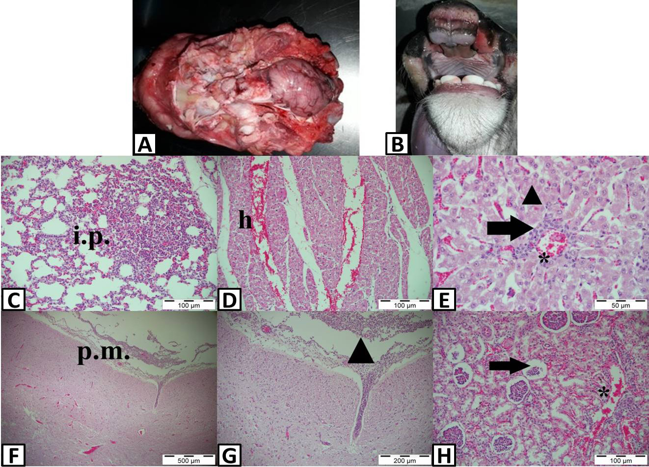
A, Anencephaly. B, Palatoschysis. C, Lung, interstitial pneumonia (i.p.), H&E, 100 μm. D, Heart, hemorrhage (h), H&E, 100 μm. E, Liver, sinusoidal hyperemia (star), mononuclear cell infiltration (arrow), fat degeneration (arrowhead), H&E, 50 μm. F, Brain, purulent meningitis (p.m and arrowhead), H&E, 500 μm. G, Higher magnification, H&E, 200 μm. H, Kidney, hemorrhage (star), atrophy and necrosis in the glomeruli (arrow), H&E, 100 μm.

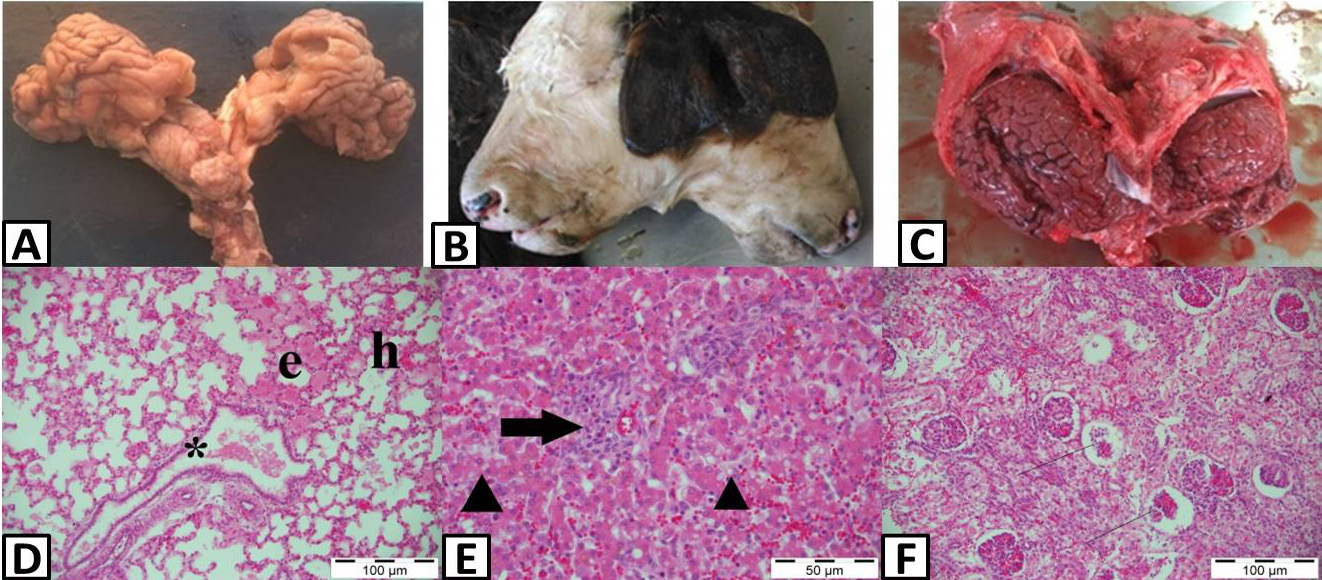
A, Diencephali. B, Diprosopus monauchenos. C, Diencephali. D, Lung, edema (e), bronchiole content (star), hyperemia (h), H&E, 100 μm. E, Liver, sinusoidal hyperemia, fat degeneration in hepatocytes (arrowheads), inflammatory cell infiltration (arrow), H&E, 50 μm. F, Kidney, hyperemia, hemorrhage, atrophy and necrosis of glomeruli (lines), H&E, 100 μm.
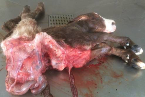
Schistosoma reflexum.


