
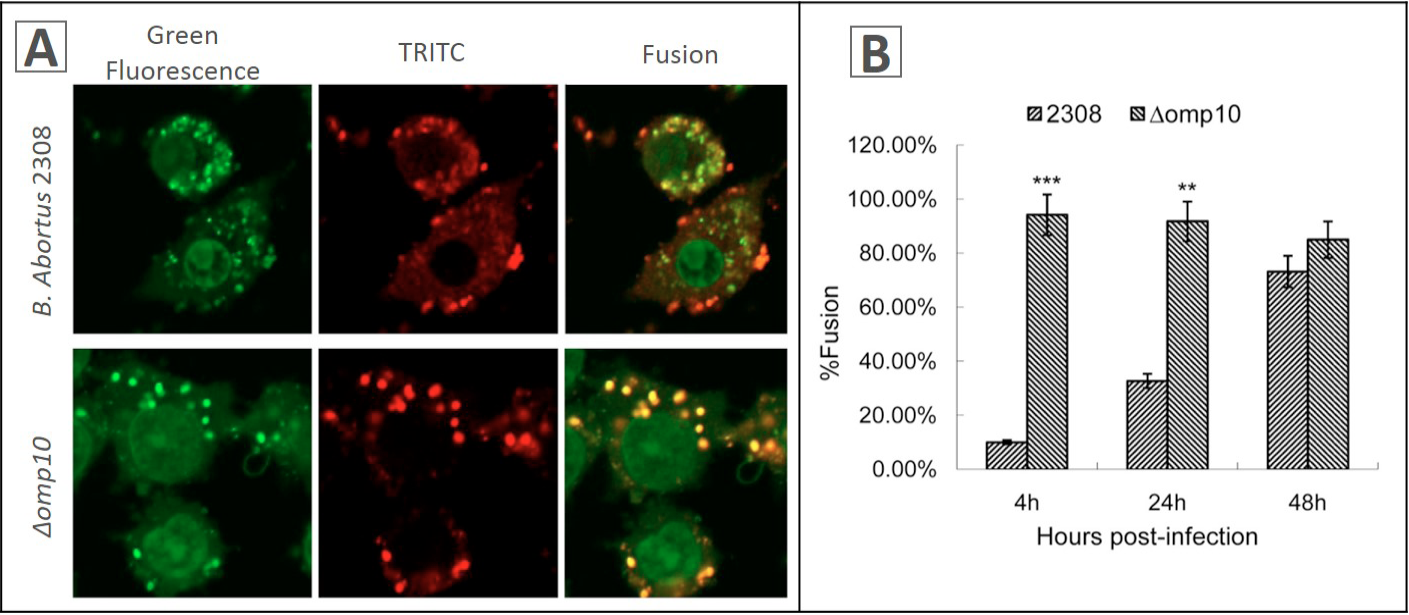
Fig 5
Interaction of B. abortus 2308- and mutants-containing phagosomes with lysosomes in RAW264.7 macrophages. B. abortus were labeled with sheep anti-B. abortus IgG antibodies and Rhodamine (TRITC) conjugated AffiniPure donkey anti-sheep IgG antibody, lysosomes were labeled with Cell NavigatorTM Lysosomes Staining Kit Green Fluorescence. A, cells were fixed at different time points after infection. Confocal images of cells containing B. abortus were obtained at 4 h post-infection; B percentage of phagosome-lysosome fusion at different time points after infection. Fusion was evaluated by the colocalization of markers, Green fluorescein and TRD. To determine the percentage of fusion, bacteria were analyzed at each time point.